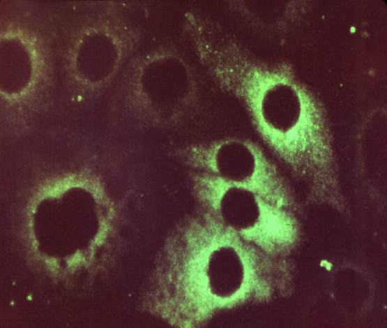
Mouse anti Interleukin-1 beta (IL-1 b) antibody co ...

Details
Clone
AS57
Storage
4ºC
Antibody is raised in
Mouse
Antibody's reacts with
Human
Protein number
see ncbi
French translation
anticorps
Latin name
Mus musculus
Conjugation
Biotinylated
Antibody's specificity
No Data Available
Application
Enzyme Immunoassay
Category
Primary Antibodies
Relevant references
no information yet
Antibody come from
Human recombinant interleukin 1 beta (hIL- 1§).
Antibody's reacts with these species
This antibody doesn't cross react with other species
Antigen-antibody binding interaction
Mouse anti Interleukin-1 beta (IL-1 b) antibody conjugated to Biotin Antibody
Other description
Provided as solution in phosphate buffered saline with 0.08% sodium azide. Protein A/G Chromatography
Description
This antibody needs to be stored at + 4°C in a fridge short term in a concentrated dilution. Freeze thaw will destroy a percentage in every cycle and should be avoided.
Gene
The Interleukin-1 family (IL-1 family) is a group of 11 cytokines, which plays a central role in the regulation of immune and inflammatory responses to infections or sterile insults. Rec. E. coli interleukin-1 for cell culture or antibody production.
Warnings
This product is intended FOR RESEARCH USE ONLY, and FOR TESTS IN VITRO, not for use in diagnostic or therapeutic procedures involving humans or animals. This datasheet is as accurate as reasonably achievable, but Nordic-MUbio accepts no liability for any inaccuracies or omissions in this information.
Test
Mouse or mice from the Mus musculus species are used for production of mouse monoclonal antibodies or mabs and as research model for humans in your lab. Mouse are mature after 40 days for females and 55 days for males. The female mice are pregnant only 20 days and can give birth to 10 litters of 6-8 mice a year. Transgenic, knock-out, congenic and inbread strains are known for C57BL/6, A/J, BALB/c, SCID while the CD-1 is outbred as strain.
Properties
If you buy Antibodies supplied by nordc they should be stored frozen at - 24°C for long term storage and for short term at + 5°C.Biotin conjugates can be detected by horseradish peroxidase, alkaline phosphatase substrates or anti biotin conjugated antibodies. Avidin and Streptavidin bind to the small biotin and are couple to HRP or AP for ELISA. To break the streptavidin Biotin bond we suggest to use a 6 molar guanidine HCl solution with acidity of pH 1.6.
Long description
Produced by activated macrophages, IL-1 stimulates thymocyte proliferation by inducing IL-2 release, B-cell maturation and proliferation, and fibroblast growth factor activity. IL-1 proteins are involved in the inflammatory response, being identified as endogenous pyrogens, and are reported to stimulate the release of prostaglandin and collagenase from synovial cells. The lack of a specific hydrophobic segment in the precursor sequence suggests that IL-1 is released by damaged cells or is secreted by a mechanism differing from that used for other secretory proteins.
Antibody's suited for
Applications: Neutralization: This antibody has been used at a 100-fold molar excess to neutralize ³95% of the biological activity of human IL-1 . Immunohistochemistry: This antibody has been used at 1-10 µg/ml to stain human IL-1 expressed in mononuclear cells, following fixation with 4% paraffin-formaldehyde in PBS for 10 minutes at 4¡C, plus 0.1% saponin 20 minutes at RT. Immunoprecipitation: This antibody has been used to immunoprecipitate >90% hIL-1 in solution using a double-antibody method. (Midgley, AR and Hepburn, MR, Methods Enzymol., 70, 266, 1980). ELISA: This antibody has been used as a solution phase antibody at 2 µg/ml in a two site ELISA with unconjugated IL-1beta antibody (Cat. No. L140M). Typically the ELISA has a working range of 0-500 pg/ml and a sensitivity of 5 pg/ml with the appropriate standard. Recommended sample volumes are 25-200 µl. Recovery of hIL-1( in human serum is typically >85% in the ELISA